
Condition
Endocrinology
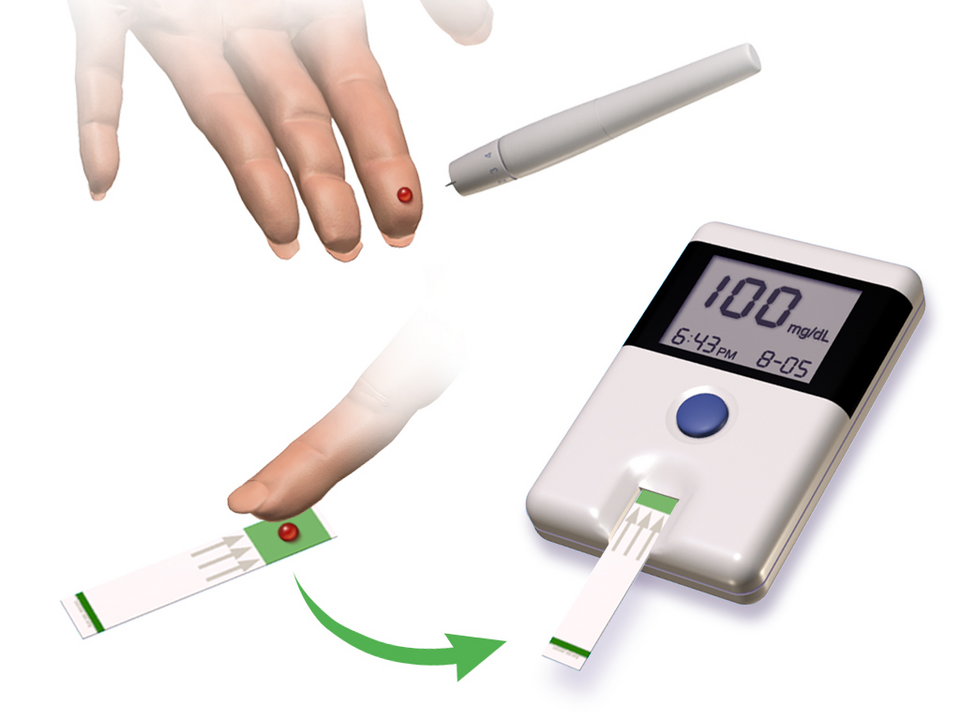
Type 2 Diabetes Mellitus
Type 2 Diabetes Mellitus is an established clinical disorder categorized under Endocrinology. Characterized by pathophysiological involvemen...
Medically ReviewedRead
Instant clinical intelligence across 310+ pharmaceutical monographs, 1,200+ verified disease guides, multi-drug interaction matrix, and evidence-based calculators.
Verified clinical reference databases, drug safety checkers, and immunization protocols.
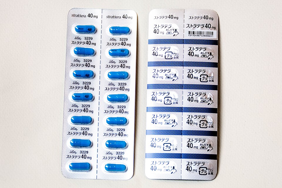
310+ verified prescription & OTC drugs, adult dosages, contraindications & interactions.
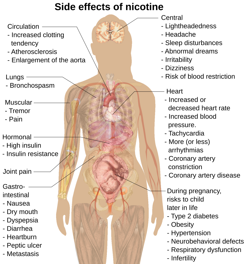
Comprehensive clinical guides covering etiology, symptoms, diagnosis & treatments.

Multi-medication safety engine detecting pairwise clinical interactions and risks.

National Immunization Schedule (NIS) timeline from birth to 16 years with catch-up rules.
Normal reference ranges, clinical indications, and interpretations for routine labs.
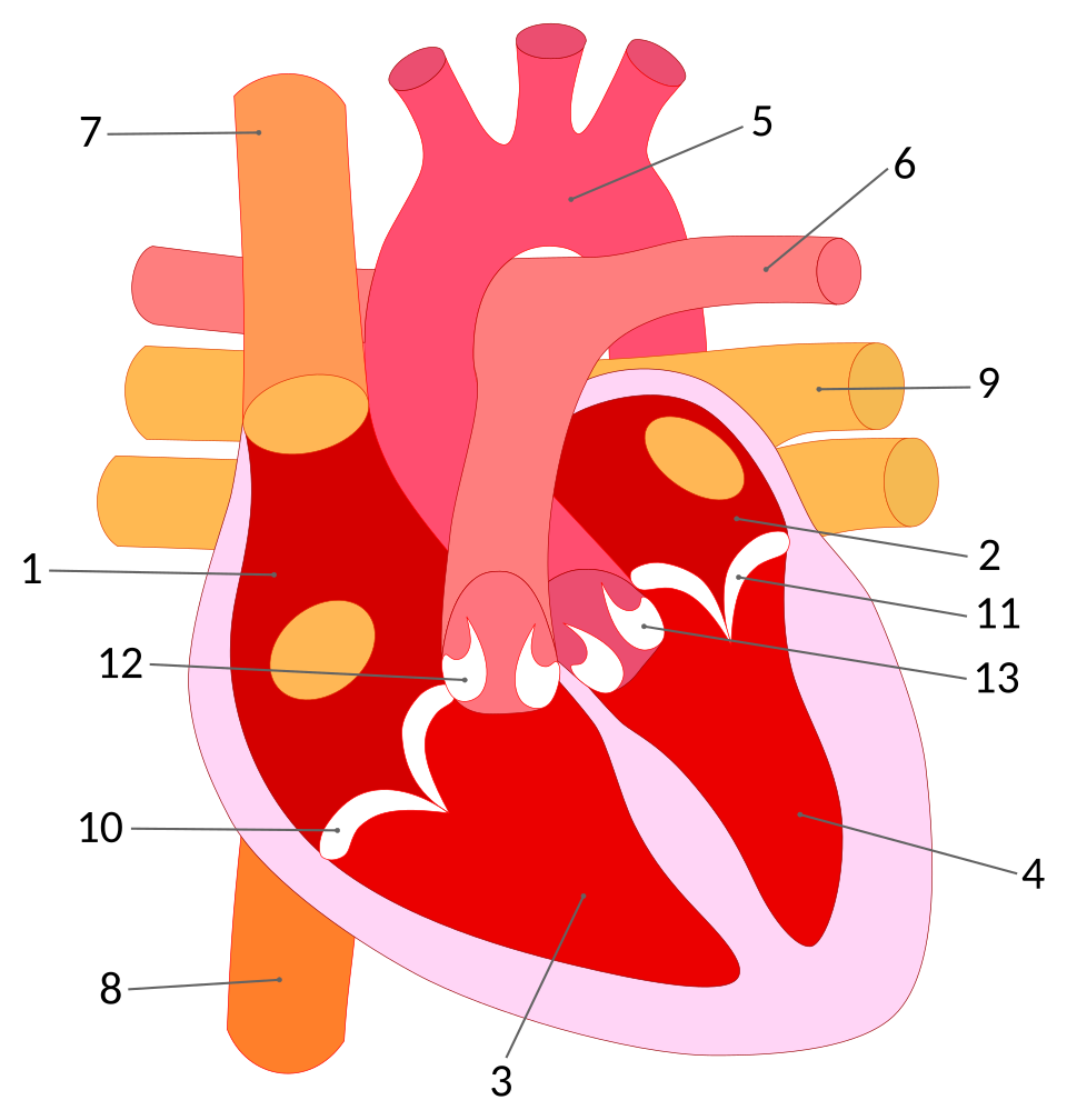
In-depth anatomical references of the cardiovascular, neural, and renal systems.
Evidence-based disease overviews, clinical symptoms, risk factors, diagnostic criteria, and management options.
Type 2 Diabetes Mellitus is an established clinical disorder categorized under Endocrinology. Characterized by pathophysiological involvemen...
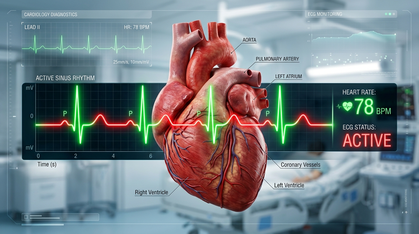
Essential Hypertension is an established clinical disorder categorized under Cardiology. Characterized by pathophysiological involvement of...
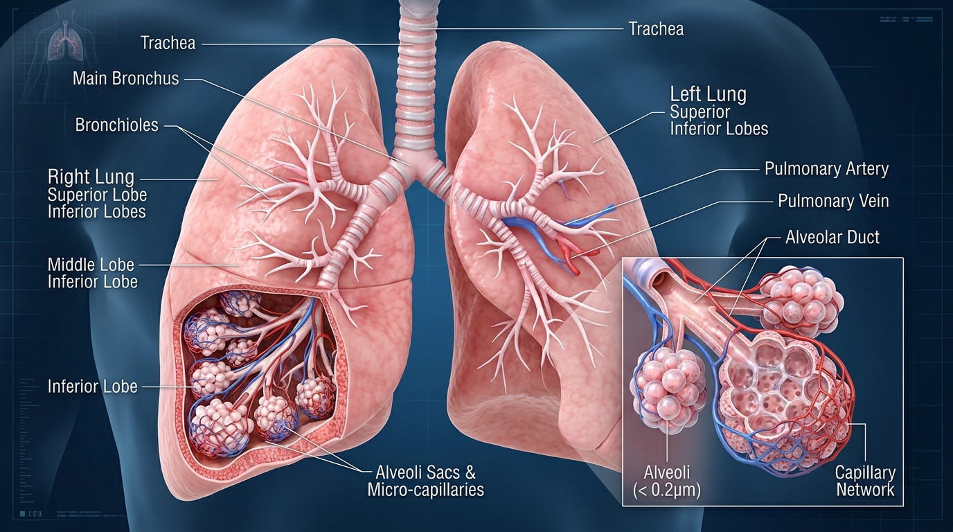
Bronchial Asthma is an established clinical disorder categorized under Pulmonology. Characterized by pathophysiological involvement of the r...

Leukemia is a broad term for malignant cancers of the body's blood-forming tissues, including the bone marrow and the lymphatic system. It i...

Ectopic pregnancy is a pregnancy in which a fertilized egg implants and begins to develop outside the normal cavity of the uterus (womb). In...

Migraine is an established clinical disorder categorized under Neurology. Characterized by pathophysiological involvement of the central ner...
Validated medical assessment tools, renal clearance estimators, ideal weight formulas, and health metrics.
Evaluate potential drug-drug interactions between multiple medications, clinical mechanisms, and precautions.
Indian National Immunization Schedule (NIS) timeline from birth to 16 years with dose numbers and catch-up rules.
Educational step-by-step symptom assessment, red-flag emergency detection, and healthcare recommendations.
We investigate popular health claims by separating what people believe from what scientific evidence actually supports.

The popular claim investigated — bromelain, mucociliary clearance, and what science actually says.
Kidney filtration, ADH hormone, thermoregulation — the real biology of hydration explained.

Vitamin D synthesis, circadian rhythm, UV damage — the complete science of sun exposure.

Probiotics, live cultures, microbiome — separating genuine science from marketing claims.
Immune defenses naturally decline past 50. Protecting yourself against preventable illnesses preserves vitality and long-term health.
As our bodies mature, waning childhood antibodies and natural immunosenescence leave adults vulnerable to debilitating conditions like shingles, pneumococcal pneumonia, and severe influenza complications.
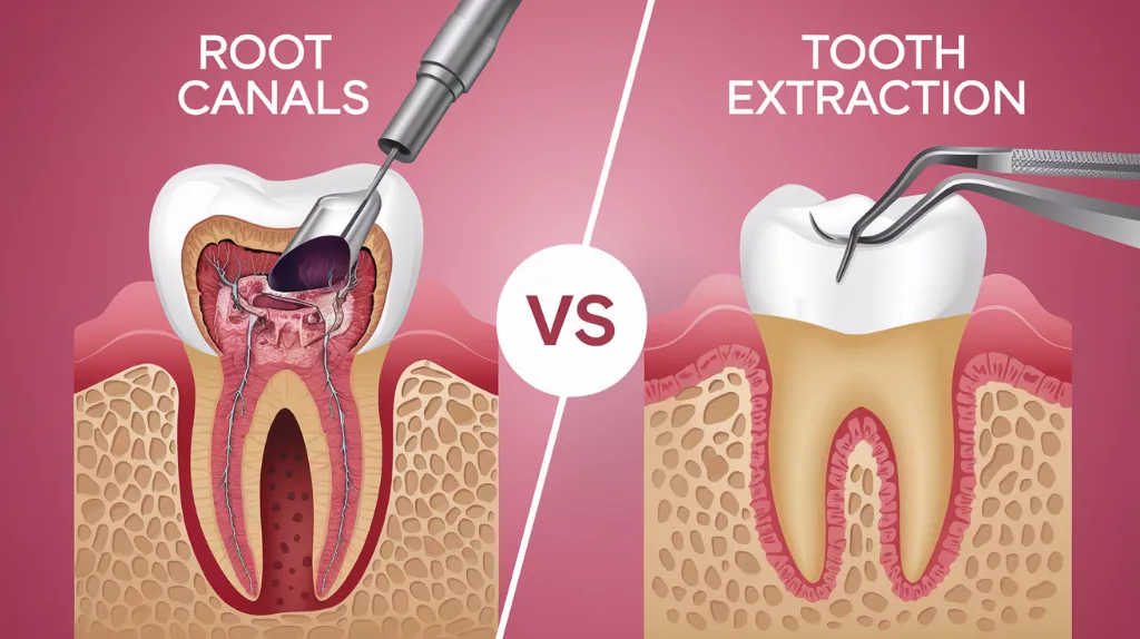
Understand the treatment that can help save a damaged or infected tooth.
Understand the symptoms, procedure, pain, recovery, aftercare, and key differences between root canal treatment and tooth extraction.
Ectopic pregnancy is a pregnancy in which a fertilized egg implants and begins to develop outside the normal cavity of the uterus (womb). In most cases, the pregnancy develops within a fallopian tube, although ectopic implantation can occur in other locations, including the cervix, ovary, or abdominal cavity.

Transvaginal Sonography • Tubal Ring Sign
High-resolution transvaginal sonography (TVUS) visualizes the empty uterine cavity, identifies the adnexal "bagel/ring" sign, detects free blood in the pouch of Douglas, and verifies live ectopic cardiac activity.
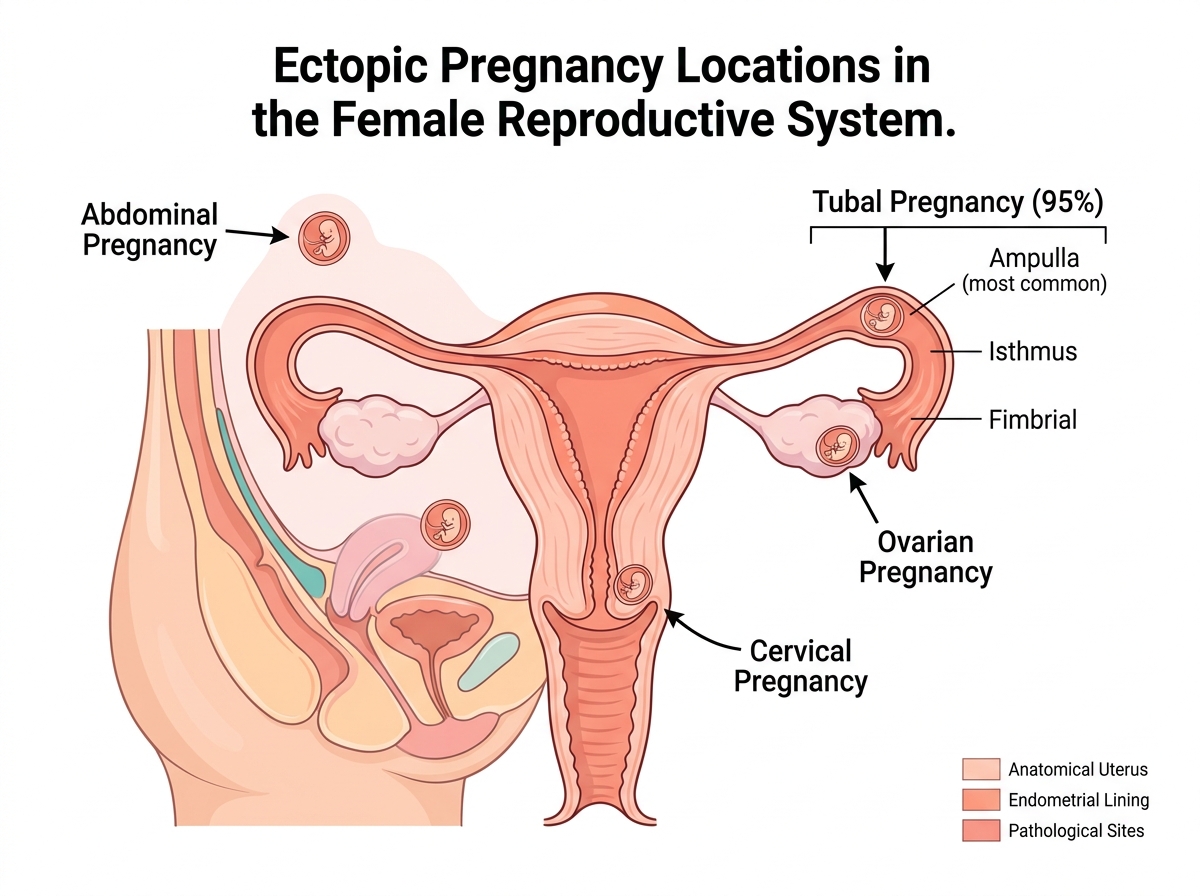
Ampulla · Isthmus · Fimbrial · Interstitial
95% implant in the fallopian tube (ampulla 70%, isthmus 12%, fimbrial 11%). The remaining 5% occur in the ovary (3%), cervix (1%), or abdominal cavity (1%).
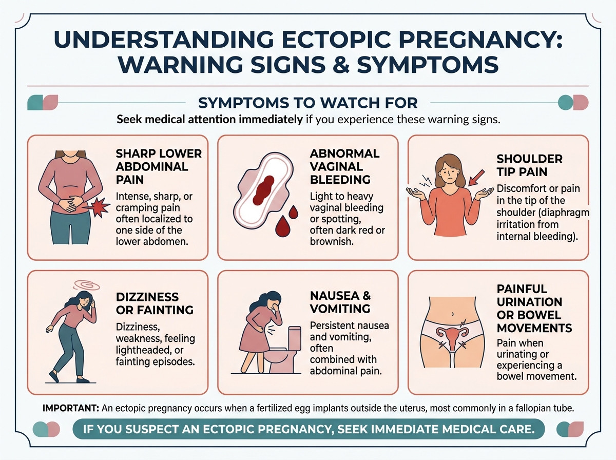
Abdominal Pain · Spotting · Shoulder Tip
Sharp unilateral lower abdominal pain, abnormal vaginal spotting, shoulder tip pain from diaphragmatic irritation, and weakness or dizziness from internal bleeding.
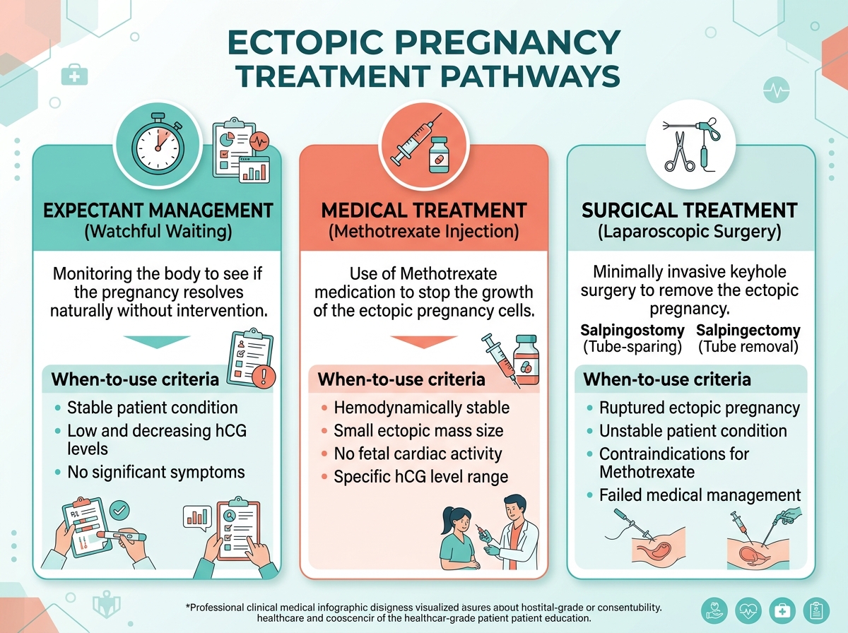
Watchful Waiting · Medical · Laparoscopy
Treatment depends on hemodynamic stability, β-hCG levels, and mass size. Expectant management, Methotrexate (88–96%), or emergency laparoscopy (salpingostomy/salpingectomy).
Clear, evidence-based medical information explaining causes, early warning signs, diagnostic standards, treatment modalities, and emergency triage.
An ectopic pregnancy may occur when the normal movement of a fertilized egg through the fallopian tube is disrupted. Factors associated with an increased risk include:
Symptoms commonly develop during the early stages of pregnancy and may include:
Diagnosis generally involves a combination of:
An ectopic pregnancy cannot develop normally outside the uterus and cannot safely be relocated to the uterus. Treatment depends on factors such as the pregnancy's location and size, β-hCG levels, symptoms, and the individual's overall condition.
Treatment may include:
A ruptured ectopic pregnancy can cause life-threatening internal bleeding. Severe abdominal or pelvic pain, fainting, marked dizziness, weakness, or shoulder-tip pain in someone who may be pregnant requires urgent emergency medical assessment.
Early diagnosis and appropriate treatment are important for protecting the patient's health and, when possible, preserving future fertility.
Under normal physiology, fertilization occurs in the ampulla of the fallopian tube. The developing blastocyst is propelled over 3 to 4 days toward the uterus by coordinated ciliary beating and smooth muscle peristalsis, implanting into the hormone-primed endometrium. In an ectopic pregnancy, this transport is arrested, and the blastocyst implants into tissues incapable of supporting placental invasion.
Prior PID, surgery, or endometriosis creates intraluminal adhesions, ciliary destruction, or structural kinking that physically traps the fertilized egg before it reaches the uterus.
Hormonal imbalances (progesterone/estrogen), smoking-induced ciliary paralysis, or tubal muscular dysfunction slow the egg's 3–4 day journey through the fallopian tube.
The blastocyst implants into the thin tubal wall, eroding into blood vessels. As trophoblast cells proliferate (doubling every 48 hrs), the inelastic tube distends and eventually ruptures.
When an Rh-negative mother carries an Rh-positive baby, fetomaternal blood mixing can trigger lifelong antibody production against fetal blood cells. Learn the essential blood tests used to check if antibodies are in the maternal bloodstream and how timely Anti-D Immunoglobulin (RhoGAM) prevents Hemolytic Disease of the Fetus and Newborn (HDFN).
Before giving Anti-D Immunoglobulin (RhoGAM), obstetricians must verify whether the mother has already been sensitized (isoimmunized). If maternal anti-D antibodies have already formed, RhoGAM cannot reverse them. The gold-standard diagnostic tool is the Indirect Coombs Test (Indirect Antiglobulin Test - IAT).
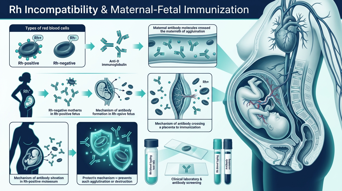
Maternal Antibody Screen / Circulating Anti-D Test
Checks whether the Rh-negative mother has ALREADY developed circulating anti-Rh (anti-D) antibodies against fetal red blood cells.
Serial Antibody Quantitation Assay
Measures the precise concentration/strength of circulating anti-D IgG antibodies in maternal blood via serial dilutions (e.g., 1:2, 1:4, 1:8, 1:16, 1:32).
Blood Group & Rh Typing (Forward & Reverse)
Determines whether the mother is Rh-negative (dd) and verifies the biological father’s Rh status (Rh-positive DD or Dd vs Rh-negative dd).
Newborn Cord Blood Antiglobulin Test
Tests whether maternal anti-D antibodies have already crossed the placenta and physically bound onto the newborn baby’s red blood cells.
Fetomaternal Hemorrhage (FMH) Quantification
Quantifies the exact volume of fetal red blood cells that have crossed into maternal circulation during delivery or traumatic events.
Non-Invasive Prenatal Testing (NIPT) for Fetal RhD
Analyzes circulating fetal DNA fragments in maternal plasma to non-invasively determine whether the unborn baby is RhD-positive or RhD-negative.
Whether you are protecting healthy kidneys or managing Chronic Kidney Disease (CKD Stages 1 through 5), precision nutrition is your most potent clinical defense. Learn how managing the 5 Core Renal Pillars—Sodium, Potassium, Phosphorus, Protein, and Fluids—protects nephrons, prevents toxic uremic buildup, and sustains energy and longevity.

8+ Clinically Verified Renal Superfoods
Blueberries deliver anthocyanin antioxidants that protect glomerular capillaries without potassium overload. Red bell peppers supply full-spectrum vitamin A, C, and B6 at only 211 mg K+ per 100g. Cauliflower provides the perfect low-potassium mashed potato alternative (176 mg K+). Extra virgin olive oil adds heart-protective oleic acid and polyphenols with zero electrolyte load.

HBV Protein · Zero Phosphorus Yolk · Rich Antioxidants
Two egg whites deliver High Biological Value (HBV) protein with virtually zero phosphorus (all egg phosphorus is in the yolk). Paired with vitamin-C-rich red and yellow bell peppers (boosts iron absorption) and antioxidant-rich blueberries (114 mg K+ / 100g). Drizzled with anti-inflammatory garlic olive oil. Estimated total: ~15g protein, <200mg sodium, ~250mg potassium.

Omega-3 Rich · Low Potassium · Anti-Inflammatory
A 3 oz grilled wild cod fillet delivers 20–24g of high-quality protein with moderate phosphorus (180mg) and heart-protective omega-3 EPA/DHA. Paired with creamy steamed cauliflower mash (176 mg K+/100g) replacing dangerous mashed potatoes, and crisp steamed green beans. Total dinner: ~24g protein, ~50mg sodium, ~480mg potassium — within Stage 3 CKD targets.
Nutrient requirements shift dramatically as glomerular filtration rate (eGFR) changes. Switch below to see precise targets.
Significant reduction in functional nephrons. Accumulation of urea nitrogen, early phosphate retention, impaired vitamin D activation, and secondary hyperparathyroidism.
Protein restriction without protein-energy wasting, monitoring serum bicarbonate (>22 mEq/L) to prevent metabolic acidosis.
Explore clinical safety profiles, electrolyte values, and exact kitchen substitutions to protect failing kidneys while maintaining gourmet flavor.
Rich in vitamin C, folate, and sulforaphane. An outstanding low-potassium alternative that can be mashed to replace high-potassium mashed potatoes.
Packed with anthocyanin antioxidants that fight systemic inflammation and protect glomerular capillaries without potassium overload.
Unlike most vegetables, red bell peppers provide abundant vitamin A, C, and B6 while maintaining an exceptionally low potassium profile.
The gold-standard High Biological Value (HBV) protein. 100% pure protein with almost zero phosphorus, as all egg phosphorus is concentrated in the yolk.
Calorie-dense healthy monounsaturated fat (oleic acid) and polyphenols that combat cardiovascular risk in renal patients without mineral load.
Contains allicin and quercetin. Delivers intense savory flavor so you can eliminate added table salt without compromising taste.
Cruciferous vegetable loaded with vitamins K and C, insoluble fiber, and phytochemicals that promote gut microbiome health and lower uremic toxins.
High in pectin, a soluble fiber that lowers cholesterol, regulates blood glucose, and reduces nitrogenous load.
A single banana delivers roughly 20% of an advanced CKD patient’s entire daily potassium budget. Can trigger sudden cardiac arrhythmia.
Severe hyperkalemia hazard if boiled whole or fried. Can only be eaten in strict moderation IF leached using the clinical 4-step boiling method.
FATAL NEUROTOXIN DANGER. Contains carambolin, an oxalate neurotoxin. Healthy kidneys clear it, but in CKD it crosses into the brain causing intractable hiccups, seizures, and death.
Loaded with inorganic phosphoric acid. Unlike organic plant phosphorus (40% absorbed), inorganic phosphate is 90–100% absorbed into the bloodstream, accelerating vascular calcification.
A triple threat for kidneys: loaded with sodium chloride (fluid retention), inorganic phosphate salts, and advanced glycation end products.
Brands like Nu-Salt or LoSalt substitute sodium with potassium chloride. One pinch can cause severe hyperkalemia and acute fatal cardiac arrest in CKD patients.
Supplies high-quality protein and omega-3 fatty acids (EPA/DHA) that lower systemic triglycerides. Must be portion-controlled (3 to 4 ounces) in Stages 3–4.
Much lower in potassium and phosphorus than cow’s milk (which contains ~380 mg K and 230 mg P per cup). Check label to confirm no added tricalcium or potassium phosphate.
Root vegetables like potatoes, carrots, yams, and squash can be made safer for CKD Stages 3–5 by leaching excess potassium ions into warm water before cooking.
Engineered for CKD Stage 3 (~1,800 kcal, ~50g protein, <1,500mg sodium, low potassium, low phosphorus).
2 egg whites scrambled in olive oil with diced red bell peppers + 1/2 cup fresh blueberries + 1 slice sourdough toast.
1 small crisp apple sliced and dusted with ground cinnamon (zero sodium, rich in soluble pectin fiber).
3 oz fresh cod with lemon dill + 1 cup steamed cauliflower mash drizzled with garlic-infused olive oil + steamed green beans.
3 oz chicken tenderloins sautéed with shredded purple cabbage, garlic, and scallions over 1/2 cup jasmine white rice.
Failing kidneys lose the ability to clear specific dietary toxins and mineral spikes. Avoid these catastrophic hazards:
NEVER eat star fruit or drink star fruit juice. It contains carambolin, an oxalate neurotoxin that accumulates in renal failure, causing seizures, coma, and permanent brain damage.
"Low sodium" salts replace sodium with potassium chloride. Just 1 teaspoon delivers over 800 mg of potassium, risking sudden fatal ventricular tachycardia.
Over-the-counter painkillers (Ibuprofen, Naproxen, Diclofenac) constrict renal afferent arterioles, precipitating acute on chronic renal failure.
Inspect ingredient lists for "Pyrophosphate", "Dicalcium phosphate", or "Phosphoric acid". Inorganic forms are 100% absorbed, petrifying blood vessels.
Every kidney is unique. Serum potassium, eGFR, proteinuria, and bicarbonate fluctuate with medication and disease etiology. Always work alongside a Registered Dietitian Nutritionist (RDN) specializing in clinical nephrology.
Expert-written health guides, clinical updates, and preventive medicine insights.

The human gastrointestinal tract houses trillions of microorganisms that produce neurotransmitters, modulate systemic immune responses, and...
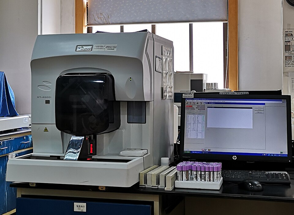
Learn the difference between daily maintenance controller inhalers and fast-acting rescue bronchodilators, common environmental triggers, an...

Not all cholesterol is harmful. Understand how Low-Density Lipoproteins (LDL) contribute to atherosclerotic plaques and how High-Density Lip...
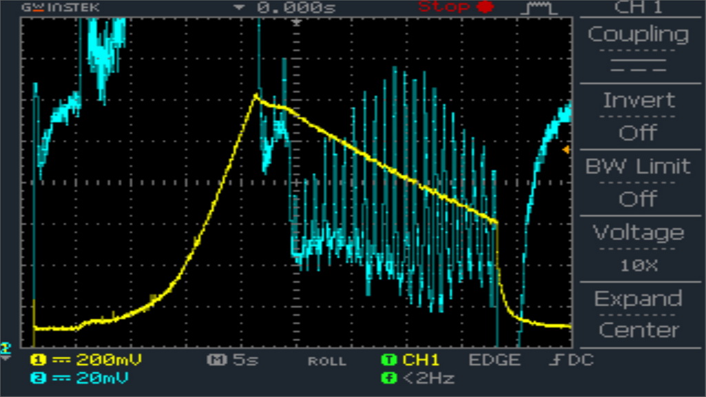
High blood pressure often exhibits no overt symptoms until irreversible vascular damage occurs. Learn how to accurately measure blood pressu...
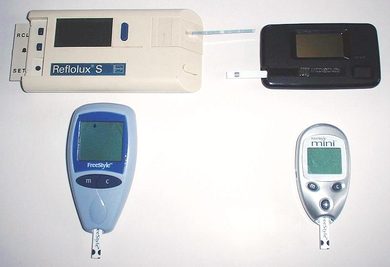
Type 2 Diabetes Mellitus is a chronic metabolic condition characterized by insulin resistance and relative insulin deficiency. Learn how a b...
Medi Info Hub follows strict clinical guidelines sourced directly from the World Health Organization (WHO), CDSCO, Indian Pharmacopoeia Commission (IPC), and national medical regulatory authorities.
Important: The information provided on this website is for educational purposes only and is not intended as a substitute for professional medical advice, diagnosis, or treatment. Always seek the advice of a qualified healthcare provider with any questions you may have regarding a medical condition.